
Meniscus Tears: Types, Diagnosis, Repair vs. Meniscectomy, and Long-Term Outcomes results
The menisci are two crescent-shaped fibrocartilaginous structures in the knee that serve essential mechanical, shock-absorbing, and stabilizing functions. Meniscus tears are among the most common knee injuries encountered in orthopedic practice, with an incidence of approximately 61 per 100,000 person-years. Partial meniscectomy is the most frequently performed orthopedic surgery in many countries. However, growing evidence highlights the critical importance of meniscus preservation to protect against accelerated osteoarthritis. This article provides a thorough, clinically grounded overview. For the official patient guide, visit AAOS — Meniscus Tears.
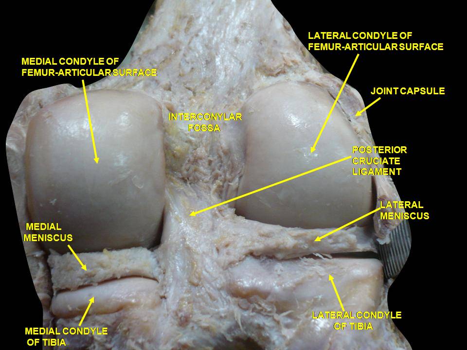
Meniscus Anatomy and Function
The medial meniscus is C-shaped, larger, and relatively fixed because its outer horn is firmly attached to the joint capsule and the deep medial collateral ligament (MCL). The lateral meniscus is more circular (nearly O-shaped), smaller, and more mobile, covering a greater proportion of the tibial plateau surface than the medial side.
Both menisci are composed of fibrocartilage — a dense extracellular matrix of type I collagen fibers (circumferentially oriented to resist hoop stresses) interspersed with water (approximately 65–75% of wet weight) and proteoglycans. This unique structure allows them to function as load distributors, shock absorbers, secondary stabilizers, and contributors to joint lubrication and proprioception.
Critically, the menisci transmit 50–70% of the compressive load across the knee joint. Loss of meniscal tissue dramatically increases contact pressures on the articular cartilage: a total meniscectomy can increase peak contact stress by up to 235% in the medial compartment — a key reason why meniscus preservation is now a central principle of modern orthopedic care.
The vascular supply of the meniscus is a crucial concept that directly influences treatment decisions. The outer third (red zone) is well-vascularized by the medial and lateral geniculate arteries and has good healing potential. The middle third (red-white zone) has variable blood supply and unpredictable healing. The inner third (white zone) is avascular and relies on synovial fluid diffusion for nutrition, making spontaneous healing virtually impossible in this region.
Classification of Meniscus Tears
Meniscus tears are classified by morphology (tear pattern), location (zone), chronicity, and etiology (traumatic vs. degenerative). Understanding the tear pattern is essential because it determines whether the tear is repairable or requires partial meniscectomy.
Tear patterns
Tear Pattern | Description | Repairability | Common Mechanism |
|---|---|---|---|
Longitudinal (vertical) | Split parallel to the circumferential fibers; runs along the long axis | High (especially in red/red-white zone) | Twisting on a loaded knee (sports) |
Radial | Full-thickness split perpendicular to the long axis; disrupts hoop stress | Low to moderate (depends on size and location) | Squatting or twisting under load |
Horizontal (cleavage) | Split within the substance, parallel to the tibial surface | Generally not repaired | Degenerative; common in older adults |
Bucket-handle | Large longitudinal tear with a displaced fragment that flips into the intercondylar notch | High (if in vascular zone); urgent if locked | Significant twisting injury; can lock the knee |
Complex | Combination of multiple patterns (e.g., radial + horizontal + longitudinal) | Low (often requires partial meniscectomy) | Usually degenerative; multi-directional degeneration |
Flap (parrot-beak) | Oblique tear with a free fragment that can catch and cause mechanical symptoms | Low to moderate | Repetitive microtrauma or acute twist |
Root tear | Detachment of the meniscus from its posterior or anterior root attachment | Repairable and should be repaired when recognized | Degenerative (medial) or acute (lateral); increasingly diagnosed |
Clinical Presentation
Patients with acute traumatic meniscus tears typically report a twisting or squatting mechanism followed by joint-line pain (medial for medial meniscus, lateral for lateral meniscus), swelling that develops over 24–48 hours (slower than ACL hemarthrosis), and mechanical symptoms such as catching, locking, or a sense of something "getting stuck" in the knee.
In contrast, degenerative meniscus tears (more common in patients over 40) often present with insidious onset of mild joint-line pain, intermittent swelling after activity, and occasional clicking — without a clear traumatic event. These tears frequently coexist with osteoarthritis, making the treatment decision more nuanced.
Physical examination typically reveals localized joint-line tenderness (sensitivity 74–88%, specificity 59–79%), a positive McMurray test (sensitivity 29–61%, specificity 77–98%), and sometimes an effusion. The Thessaly test (pain with standing rotations at 5° and 20° flexion) has shown promising sensitivity (66–89%) in some studies.
It is essential to also evaluate for concurrent ligament instability (especially ACL integrity, since ~50% of ACL tears have associated meniscal injuries), range of motion limitations, and any mechanical block to full extension or flexion that might indicate a displaced bucket-handle fragment requiring urgent surgical attention.
Diagnostic Workup
MRI is the imaging modality of choice for meniscus evaluation, with sensitivity of 77–95% and specificity of 73–98%. On MRI, a meniscus tear appears as high signal intensity extending to at least one articular surface on T2-weighted or proton-density fat-suppressed sequences. Grade 1 (intrameniscal signal not reaching a surface) and Grade 2 (signal reaching one surface but not both) are generally not considered true tears, while Grade 3 (signal reaching both articular surfaces) confirms a full-thickness tear.
Imaging decision framework
function meniscusImagingDecision(patient):
  exam = performKneeExam()

  if exam.mechanicalLocking or exam.blockedExtension:
    // Locked knee — urgent MRI + likely surgical referral
    orderUrgentMRI()
    referOrthopedicsUrgent()
    return "URGENT"

  if exam.jointLineTenderness AND exam.positiveMcMurray:
    // High clinical suspicion
    orderMRI()
    return "STANDARD_WORKUP"

  if patient.age > 40 AND exam.mildJointLinePain AND noMechanicalSymptoms:
    // Likely degenerative; consider trial of conservative care first
    // MRI may not change initial management
    considerConservativeCareFirst()
    if noImprovementAfter6to12Weeks():
      orderMRI()
    return "CONSERVATIVE_FIRST"

  if exam.suggestsACLinjury:
    // MRI to evaluate both ACL and meniscus
    orderMRI()
    return "COMBINED_WORKUP"

  return "INDIVIDUALIZE"
Treatment: When to Repair, When to Resect, and When to Rehabilitate
The treatment paradigm for meniscus tears has shifted significantly over the past two decades. The old approach of routine partial meniscectomy ("just clean it up") has been replaced by a meniscus-preserving philosophy grounded in long-term outcome data.
Partial meniscectomy outcomes — the evidence shift
A landmark 2013 randomized trial in the New England Journal of Medicine (Sihvonen et al.) compared arthroscopic partial meniscectomy to sham surgery for degenerative meniscus tears and found no significant difference in outcomes at 2 years. Subsequent trials and meta-analyses have largely confirmed that for degenerative meniscus tears without mechanical locking, surgery offers no advantage over structured physical therapy. This has led major guidelines (including from the AAOS and the Danish Health Authority) to recommend non-surgical management as first-line for most degenerative tears.
However, for traumatic tears in young, active patients — especially repairable longitudinal tears in the vascular zone — meniscus repair remains the standard of care, with success rates of 60–90% depending on tear pattern, zone, and surgical technique.
Treatment decision matrix
Patient Profile | Tear Characteristics | Recommended Approach | Rationale |
|---|---|---|---|
Young athlete (<35) | Acute longitudinal tear, red/red-white zone | Meniscus repair | Preserve tissue; high healing potential; prevent OA |
Young athlete (<35) | Displaced bucket-handle causing locking | Urgent meniscus repair | Relieve locking; preserve meniscus; prevent chondral damage |
Any age | Root tear (medial or lateral) | Root repair (if no advanced OA) | Root detachment = functional meniscectomy; repair restores hoop stresses |
Adult 40–60 | Degenerative tear, no mechanical symptoms | Physical therapy first | NEJM-grade evidence: surgery no better than PT for degenerative tears |
Adult 40–60 | Degenerative tear WITH persistent locking/catching after 3+ months of PT | Consider partial meniscectomy | Failed conservative care; mechanical symptoms suggest flap or displaced fragment |
Any age | Complex, degenerative, white-zone tear | Minimal partial meniscectomy (if surgery needed) or PT | Not repairable; remove only the unstable fragment; preserve as much tissue as possible |
Rehabilitation After Meniscus Surgery
Rehabilitation protocols differ significantly between meniscus repair and partial meniscectomy, reflecting the different healing demands of each procedure.
After meniscectomy: Weight-bearing as tolerated (WBAT) from day one; early range of motion; return to light activities within 1–2 weeks; return to sport typically at 4–6 weeks. Rehab focuses on quadriceps and hip strengthening, swelling control, and gradual activity progression.
After meniscus repair: More protective protocol — typically toe-touch weight-bearing (TTWB) or non-weight-bearing (NWB) for 4–6 weeks with a hinged brace; limited flexion (often 0–90° for medial repairs, 0–70° for lateral repairs) to protect the repair site; no deep squatting or twisting for 8–12 weeks; return to sport at 3–4 months. The more restrictive protocol reflects the need for the repaired tissue to heal, which is a slower biological process than recovery from meniscectomy.
Meniscus Root Tears: An Emerging Understanding
Meniscus root tears — detachments of the meniscus from its bony attachment at the posterior or anterior horn — represent a relatively newly recognized but clinically important subset of meniscus injuries. The posterior medial meniscus root tear (PMMRT) is the most common type and is strongly associated with rapidly progressive medial compartment osteoarthritis because root detachment eliminates the meniscus's hoop stress function — effectively creating a functional total meniscectomy despite the meniscus body being intact.
Diagnosis requires a high index of suspicion and specific MRI criteria (extrusion of the meniscus body >3 mm beyond the tibial margin, root discontinuity, and bone marrow edema at the root attachment). When recognized and treated with transtibial pull-out repair before significant OA develops, outcomes are encouraging. Delayed diagnosis — unfortunately common — often means the window for joint-preserving surgery has closed.
Long-Term Outcomes and Osteoarthritis Risk
The long-term consequences of meniscus loss are well documented. After total meniscectomy, the rate of radiographic OA is 50–80% at 10–20 years. Even partial meniscectomy carries an increased OA risk, though it is lower than total meniscectomy. In contrast, successful meniscus repair appears to provide better long-term joint preservation, with lower rates of OA at 10–15 year follow-up compared with meniscectomy.
Preserve meniscus tissue whenever biologically and mechanically feasible.
Not all meniscus tears need surgery. Degenerative tears without mechanical symptoms should start with rehabilitation.
Locked knees need urgent evaluation. A displaced bucket-handle can irreversibly damage cartilage within days.
Think about root tears. Unexplained rapid medial compartment OA or meniscal extrusion on MRI should raise suspicion.
Long-term monitoring matters. Even after successful treatment, patients with meniscus injuries should be monitored for early OA signs.
Educational content only — not medical advice. If you experience knee locking, persistent joint-line pain, swelling after activity, catching, or giving way, seek in-person orthopedic evaluation. Early and accurate diagnosis of meniscus tears — especially repairable ones — can significantly impact long-term joint health. Resources: AAOS Meniscus Tears, PubMed for current evidence.

