
ACL Injury: Complete Guide to Anatomier, Diagnosis, Treatment, and Return to Sports
The anterior cruciate ligament (ACL) is one of the four major ligaments that stabilize the knee joint. It is the most commonly injured knee ligament, with an estimated 200,000 ACL reconstructions performed annually in the United States alone. These injuries predominantly affect young, active individuals aged 15–35, and they carry significant short- and long-term implications for knee function, athletic participation, and joint health. This article provides a comprehensive, evidence-based overview of ACL injuries from mechanism through full recovery. For the official patient education guide, see AAOS — ACL Injuries.
Anatomy and Biomechanics of the ACL
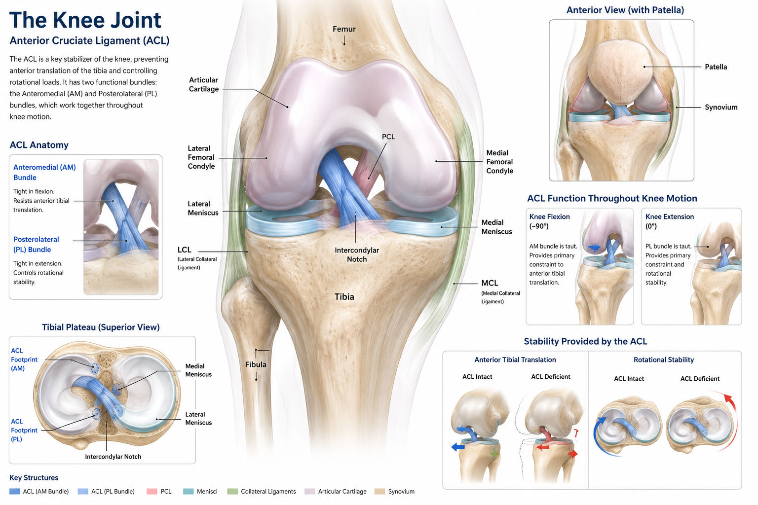
The ACL originates from the posterolateral aspect of the lateral femoral condyle and inserts onto the anterior aspect of the tibial plateau, just anterior and lateral to the intercondylar eminence (tibial spine). It measures approximately 31–38 mm in length and 10–12 mm in width, making it one of the smallest yet most critical stabilizers of the knee.
The ACL is functionally divided into two bundled fascicles: the anteromedial (AM) bundle, which is taut in flexion, and the posterolateral (PL) bundle, which is taut in extension. Together, these bundles resist anterior tibial translation, rotational instability, and combined valgus-external rotation forces.
Its primary blood supply comes from the middle geniculate artery, a branch of the popliteal artery, which enters through the intercondylar notch. The poor vascularity of the intra-articular portion is one reason why the ACL has limited intrinsic healing capacity after a complete tear.
The ACL also contains mechanoreceptors (Ruffini endings, Pacinian corpuscles) that provide proprioceptive feedback to the central nervous system. This sensory role means that an ACL-deficient knee not only loses mechanical stability but also suffers from impaired neuromuscular control, contributing to episodes of giving way even during routine activities.
Epidemiology and Risk Factors
ACL injuries account for approximately 50% of all knee ligament injuries. About 70% occur during sports participation, with the highest-risk activities being soccer, basketball, football, skiing, and gymnastics. A particularly important statistic is that female athletes are 2 to 8 times more likely to sustain an ACL injury than their male counterparts in the same sport — a disparity attributed to anatomical, hormonal, and neuromuscular factors.
Modifiable risk factors
Neuromuscular imbalances: Quadriceps dominance (relative weakness of hamstrings), valgus knee collapse during landing and cutting, and poor trunk control.
Landing mechanics: Landing with the knee extended and in valgus (knock-kneed) rather than flexed and neutral.
Insufficient neuromuscular training: Lack of proprioceptive, balance, and plyometric conditioning before sports seasons.
Fatigue: Higher injury rates are observed late in games and practices when neuromuscular control declines.
Playing surface and footwear: Artificial turf and shoe-surface interaction may increase rotational torque at the knee.
Non-modifiable risk factors
Anatomic factors: Narrow intercondylar notch width (notch stenosis), increased posterior tibial slope, and generalized joint laxity.
Sex: Wider pelvis leading to increased Q-angle, hormonal effects on ligament laxity (estrogen and relaxin), and differences in muscle activation patterns.
Genetic predisposition: Family history of ACL injury increases individual risk.
Mechanism of Injury
Approximately 70% of ACL injuries are non-contact — meaning they occur without direct impact to the knee. The classic mechanisms include:
Deceleration with cutting or pivoting: The foot is planted, the body changes direction, and the knee undergoes valgus stress combined with external tibial rotation.
Landing from a jump: Especially with the knee in near-full extension and poor hip and trunk control allowing dynamic valgus.
Direct contact: A blow to the lateral knee forcing valgus collapse (more common in football and rugby).
Skiing injuries: The "phantom foot" mechanism where the skier's arm falls back, the uphill ski is unweighted, and rotational forces tear the ACL.
Clinical Presentation
Patients almost universally report hearing or feeling a "pop" at the moment of injury. This is followed by rapid onset of a large hemarthrosis (blood in the joint) within 2–4 hours, caused by tearing of the ACL's vascular synovial covering and possibly associated meniscal or capsular bleeding.
Most athletes are unable to continue play due to pain, a sense of instability, and swelling. Some individuals, however, may walk off the field and only later develop recurrent episodes of the knee "giving way" during twisting or pivoting activities.
On examination, the knee typically demonstrates a positive Lachman test (increased anterior translation of the tibia on the femur with the knee in 20–30° of flexion), which is the single most sensitive clinical test for acute ACL rupture, with sensitivity of 85% and specificity of 94%.
Additional examination findings may include a positive anterior drawer test, a positive pivot shift test (more relevant in chronic settings), tenderness along the joint line (suggesting associated meniscal injury in up to 50% of cases), and limited range of motion due to the hemarthrosis. A thorough neurovascular exam is mandatory to rule out associated injuries.
Clinical decision algorithm
function evaluateSuspectedACL(patient):
  history = obtainMechanismAndSymptoms()
  exam = performKneeExam()  // Lachman, ADT, Pivot Shift, McMurray

  if exam.lachmanPositive AND history.popHeard:
    likelihood = "HIGH"
  elif exam.lachmanPositive:
    likelihood = "MODERATE-HIGH"
  else:
    likelihood = "LOW-MODERATE"

  if likelihood in ["HIGH", "MODERATE-HIGH"]:
    orderMRI()  // Gold standard: sensitivity 92-100%, specificity 85-100%
    assessForAssociatedInjuries()  // Meniscus (50%), MCL (30%), chondral
    discussTreatmentOptions()
  else:
    considerAlternativeDiagnosis()
    mayObserveAndReassessIn2Weeks

  return treatmentPlan
Imaging and Diagnosis
MRI is the gold standard for confirming an ACL tear and identifying associated injuries. It has a sensitivity of 92–100% and specificity of 85–100%. Typical MRI findings include discontinuity of the ligament, increased signal intensity on T2-weighted sequences, a "buckled" PCL (secondary sign), and bone bruise patterns involving the lateral femoral condyle and posterolateral tibial plateau (the classic "kissing contusion" pattern of pivot-shift injury).
Diagnostic accuracy comparison
Diagnostic Method | Sensitivity | Specificity | Clinical Role |
|---|---|---|---|
Lachman Test | 85% | 94% | Best single clinical test |
Anterior Drawer Test | 62–92% | 55–91% | Useful in chronic/subacute settings |
Pivot Shift Test | 48–73% | 95–99% | Highly specific; detects rotational instability |
MRI | 92–100% | 85–100% | Gold standard; also identifies associated injuries |
Plain X-rays | Low for ACL | N/A | Rules out fractures, avulsion fractures (Segond), OA |
Ultrasound | Limited | Limited | Not routinely recommended for ACL evaluation |
Treatment Options: Conservative vs. Surgical
Not every ACL tear requires surgery. The decision between non-operative management and ACL reconstruction depends on multiple factors: the patient's activity level, sports demands, degree of instability, associated injuries (meniscus, other ligaments), and willingness to modify activities.
Non-operative management may be appropriate for patients who are willing to give up pivoting and cutting sports, have no or minimal instability during daily activities, and are committed to a structured rehabilitation program. However, it is important to note that without an ACL, approximately 50% of patients develop meniscal tears within 5 years, which can accelerate joint degeneration.
Surgical reconstruction is generally recommended for young, active patients who want to return to pivoting sports, those with recurrent giving-way episodes, and those with associated repairable meniscal tears. The goal of surgery is not to "replace" the native ACL but to provide a stable, functional scaffold that restores sufficient stability for the patient's desired activity level.
Timing of surgery matters. Early reconstruction (within 3–5 days) is associated with a higher risk of arthrofibrosis (stiffness). Most surgeons recommend waiting 2–6 weeks after injury to allow the acute inflammation and hemarthrosis to resolve, restore preoperative range of motion (especially full extension), and begin early strengthening. Exceptions include associated injuries requiring urgent intervention, such as displaced bucket-handle meniscal tears locking the knee.
Graft choices for ACL reconstruction
Graft Type | Source | Advantages | Considerations |
|---|---|---|---|
BTB (Bone-Patellar Tendon-Bone) | Autograft (patient's own patellar tendon) | Gold standard fixation; bone-to-bone healing; fastest graft incorporation | Anterior knee pain; risk of patellar fracture; kneeling discomfort |
Hamstring (ST/G) | Autograft (semitendinosus and gracilis tendons) | Smaller incision; less anterior knee pain; good for skeletally immature patients | Slower incorporation; potential hamstring weakness; slightly higher rerupture rate in some studies |
Quadriceps Tendon | Autograft (partial quadriceps tendon with or without bone block) | Thick graft; growing evidence of excellent outcomes; good revision option | Less commonly used; surgical technique learning curve |
Allograft (Cadaveric) | Donor tissue (tibialis anterior, Achilles, BTB) | No donor site morbidity; shorter surgery; useful in multi-ligament and revision cases | Slower incorporation; higher failure rate in young active patients; disease transmission risk (extremely rare) |
Post-Operative Rehabilitation Protocol
Rehabilitation after ACL reconstruction is arguably as important as the surgery itself. A poorly executed rehab program can lead to stiffness, muscle atrophy, graft failure, or failure to return to sport. Modern protocols are criterion-based rather than time-based, though typical timelines provide a general framework.
Phase-by-phase rehabilitation
Phase | Timeline | Goals | Key Interventions |
|---|---|---|---|
Acute Protection | Weeks 0–2 | Protect graft; reduce swelling; achieve full extension; activate quadriceps | Cryotherapy; elevation; SLR exercises; heel slides; patellar mobilization; WBAT or TTWB per protocol |
Early Rehab | Weeks 2–6 | Restore flexion to 110–120°; normalize gait; improve quad control | Progressive ROM; closed-chain exercises; bike; pool therapy; gait training without crutches |
Strengthening Phase | Weeks 6–12 | Build lower extremity strength; restore proprioception; begin jogging | Leg press; step-ups; balance training; aquatic running; progressive resistance |
Dynamic Strengthening | Months 3–6 | Advance to running, agility, and sport-specific drills | Straight-line running (3–4 months); cutting/pivoting (5+ months); plyometrics; sport-specific drills |
Return to Sport | Months 9–12+ | Meet RTS criteria; pass strength and functional testing; psychological readiness | Sport-specific training; strength testing (≥90% limb symmetry); hop tests; psychological readiness questionnaires |
Return to Sport Criteria
Historically, athletes returned to sport at 6 months post-op, but mounting evidence shows this is often too early. Current consensus recommends a minimum of 9 months, with return only when all criteria are met, not simply when the calendar says so. A landmark 2016 study in the British Journal of Sports Medicine demonstrated that for every month return to sport was delayed beyond 6 months (up to 9 months), the risk of graft rerupture decreased by 51%.
Key return-to-sport criteria
Limb symmetry index (LSI) ≥ 90% on quadriceps and hamstring strength testing (isokinetic dynamometry).
Hop test symmetry ≥ 90% on single-hop, triple-hop, crossover-hop, and 6-meter timed hop.
Full range of motion (symmetrical to contralateral knee).
No effusion (no joint swelling before or after activity).
Psychological readiness: ACL-RSI (Return to Sport after Injury) score ≥ 56/100.
Sport-specific functional testing: Able to perform sport-specific drills at full speed without hesitation or compensation.
ACL Injury Prevention Programs
Multiple meta-analyses have demonstrated that neuromuscular training programs reduce ACL injury risk by 52–67%. The most well-studied programs include FIFA 11+, the PEP Program (Prevent Injury and Enhance Performance), and the Knock-Knee (KK) Program. These programs share common elements:
Biofeedback on landing mechanics: Training athletes to land with knees over toes, avoiding dynamic valgus collapse.
Hamstring and gluteal strengthening: Addressing the quad-dominant firing pattern that contributes to anterior tibial translation.
Core and trunk stability: Reducing lateral trunk lean that increases knee abduction moment.
Proprioception and balance training: Single-leg stance, wobble board, and agility drills on varied surfaces.
Plyometric training: Progressing from double-leg to single-leg jumping with proper technique.
Implementation: Programs should be performed 2–3 times per week during the preseason and maintained at least once per week during the competitive season for sustained protective effect.
Long-Term Prognosis and Osteoarthritis Risk
Regardless of whether the patient chooses operative or non-operative treatment, the long-term risk of developing knee osteoarthritis (OA) is significant. Studies report OA rates of 50–90% at 10–20 years post-injury. This is driven by the initial bone bruise (chondral damage at the time of injury), meniscal loss (if meniscectomy was performed), and altered joint biomechanics. This underscores the importance of prevention, early and optimal treatment, and long-term joint health monitoring even after successful recovery.
Key takeaways at a glance
Topic | Critical Point |
|---|---|
Incidence | ~200,000 reconstructions/year in the US; 70% sports-related |
Sex difference | Females 2–8× higher risk; neuromuscular factors are modifiable |
Best clinical test | Lachman test (85% sensitivity) |
Gold standard imaging | MRI (92–100% sensitivity); X-ray to rule out fractures |
Associated injuries | Meniscus tear in ~50%, MCL injury in ~30% |
Prevention efficacy | Neuromuscular programs reduce risk by 52–67% |
Return to sport timing | Minimum 9 months; criterion-based, not time-based |
Long-term OA risk | 50–90% at 10–20 years regardless of treatment choice |
Educational content only — not medical advice. If you experience knee instability, a pop with swelling, inability to bear weight, or any red-flag symptoms (severe deformity, numbness, weakness, fever, or loss of pulse), seek in-person orthopedic evaluation promptly. Early assessment improves outcomes. Additional resources: AAOS OrthoInfo, PubMed for research literature.

