
Kneel and Shoulders Arthroscopy: Procedures, Recovery Timelines, and Modern Rehabilitation
Arthroscopy — from the Greek arthro (joint) and skopein (to look) — is a minimally invasive surgical technique that allows orthopedic surgeons to visualize, diagnose, and treat conditions inside a joint through small incisions called portals. Since its modern refinement in the 1970s and 1980s, arthroscopy has become the most commonly performed orthopedic surgical procedure worldwide, with knee and shoulder arthroscopy representing the vast majority of cases. This article provides a thorough overview of what arthroscopy involves, common procedures, recovery expectations, and modern rehabilitation principles. For the official patient overview, see AAOS — Arthroscopy.
What Is Arthroscopy? The Technical Basics
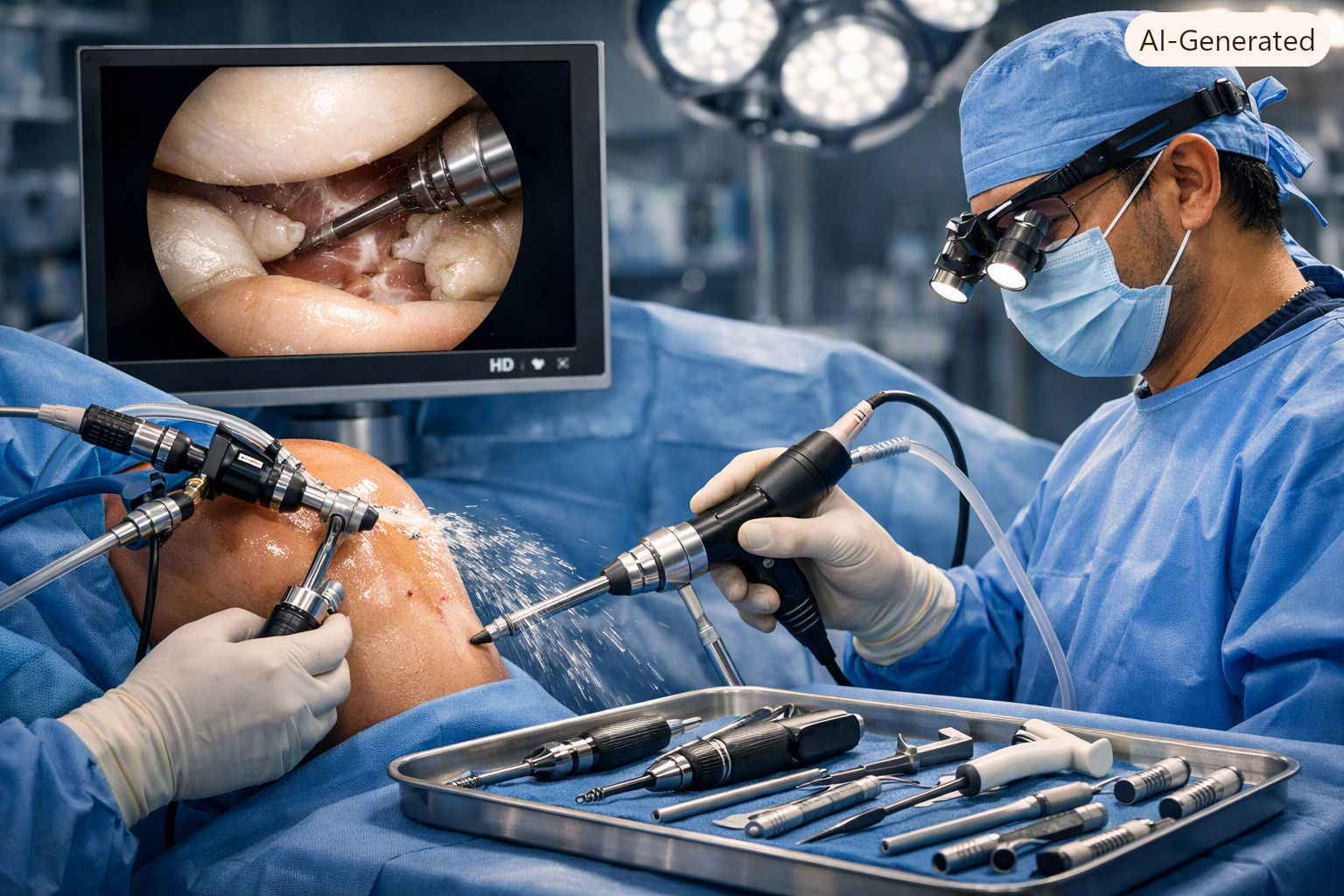
An arthroscope is a thin, fiber-optic tube (typically 2.7–4.0 mm in diameter for adult joints) equipped with a high-definition camera and a light source. The surgeon inserts it into the joint through a portal (a small incision, usually 5–10 mm), after first distending the joint with sterile saline fluid to create working space and improve visualization.
A second (and sometimes third) portal is created for working instruments — including probes, shavers, biters, thermal devices, and suture passers — that allow the surgeon to perform a wide range of procedures without making a large open incision. The entire procedure is viewed on a high-definition monitor in real time.
Common anesthesia options include general anesthesia, regional anesthesia (spinal or epidural block for lower extremity, interscalene block for shoulder), or a combination. Many patients also receive a local anesthetic injection into the joint at the end of the procedure for post-operative pain relief (intra-articular bupivacaine or ropivacaine).
The advantages of arthroscopy over traditional open surgery include smaller incisions, less post-operative pain, lower infection rates, shorter hospital stays (most are outpatient/day-case procedures), faster initial recovery, and better cosmetic outcomes. However, it is important to understand that recovery time depends primarily on what was done inside the joint, not the size of the incisions — a meniscus repair through two 5-mm portals still requires 3–4 months of protected rehabilitation.
Common Knee Arthroscopy Procedures
Knee arthroscopy accounts for approximately 60–70% of all arthroscopic procedures. The most common indications include:
Indications and typical recovery
Procedure | Indication | Weight-Bearing | Typical Recovery to Light Activity | Return to Sport |
|---|---|---|---|---|
Diagnostic arthroscopy | Unexplained pain, swelling, or mechanical symptoms when MRI is inconclusive | WBAT immediately | 1–2 weeks | 2–4 weeks |
Partial meniscectomy | Symptomatic meniscus tear not amenable to repair (degenerative, complex, white-zone) | WBAT immediately | 2–3 weeks | 4–6 weeks |
Meniscus repair | Repairable traumatic tear in vascular zone (longitudinal, bucket-handle, root) | TTWB/NWB for 4–6 weeks | 6–8 weeks | 3–4 months |
ACL reconstruction | ACL rupture in active patient desiring return to pivoting sports | WBAT with brace; protocol-dependent | 6–8 weeks (jogging at 3–4 months) | 9–12 months (criterion-based) |
Loose body removal | Free-floating cartilage or bone fragments causing locking or catching | WBAT immediately | 1–2 weeks | 2–4 weeks |
Chondroplasty / microfracture | Full-thickness cartilage defect (ICRS grade 3–4); microfracture stimulates fibrocartilage repair | WBAT → NWB/TTWB for 6–8 weeks (if microfracture) | 8–12 weeks | 4–6 months |
Lateral release | Patellar maltracking with tight lateral retinaculum (less commonly performed now) | WBAT immediately | 2–4 weeks | 6–8 weeks |
Synovectomy | Recurrent hemarthrosis, synovial chondromatosis, localized pigmented villonodular synovitis (PVNS), or inflammatory synovitis | WBAT; may require a short period of immobilization | 2–4 weeks | 4–8 weeks |
Common Shoulder Arthroscopy Procedures
Shoulder arthroscopy has expanded dramatically over the past two decades and now encompasses procedures that previously required large open incisions. The patient is typically placed in either the beach chair position (semi-sitting, like a reclined deck chair) or the lateral decubitus position (lying on the side), depending on surgeon preference and the specific procedure.
Rotator cuff repair: Reattaching a torn supraspinatus, infraspinatus, or subscapularis tendon to its footprint on the greater or lesser tuberosity using suture anchors. This is one of the most common and technically demanding arthroscopic procedures. Recovery typically involves a 4–6 week period of sling immobilization, followed by 4–6 months of progressive rehabilitation, with return to overhead sport or heavy work at 6–9 months.
Labral repair (SLAP, Bankart): The labrum is a fibrocartilaginous ring that deepens the glenoid socket. SLAP tears (Superior Labrum Anterior to Posterior) affect the top of the labrum and are common in overhead athletes. Bankart lesions involve the anteroinferior labrum and are associated with anterior shoulder dislocation. Repair involves suture anchor fixation. Sling immobilization for 4–6 weeks; return to sport at 4–6 months for SLAP, 4–6 months for Bankart (contact athletes may need longer).
Subacromial decompression (acromioplasty): Removal of bone spurs and inflamed bursa from the subacromial space to relieve impingement. Note: the evidence for isolated arthroscopic acromioplasty without rotator cuff repair has been questioned in recent years, and it is now performed less frequently as a standalone procedure.
Frozen shoulder release (capsular release): For adhesive capsulitis that has not responded to conservative treatment. The surgeon releases the thickened, inflamed capsule arthroscopically, followed by aggressive early motion. Can dramatically accelerate recovery compared to non-operative treatment alone.
Biceps tenodesis or tenotomy: Treating painful biceps tendonitis or SLAP tears by detaching the long head of the biceps from the labrum and either releasing it (tenotomy) or reattaching it to the humerus (tenotomy). Often performed as part of a larger arthroscopic procedure.
Shoulder arthroscopy recovery comparison
Immediate (aggressive)
Procedure | Sling Duration | Passive ROM | Active ROM | Strengthening Begins | Return to Sport/Work |
|---|---|---|---|---|---|
Rotator cuff repair | 4–6 weeks | Immediate (gentle) | Weeks 4–6 | Weeks 6–8 | 6–9 months |
SLAP repair | 4–6 weeks | Immediate (limited) | Weeks 4–6 | Weeks 8–12 | 4–6 months (overhead athletes: 6–9 months) |
Bankart repair | 4–6 weeks | Immediate (limited ER) | Weeks 4–6 | Weeks 6–8 | 4–6 months (contact: 6+ months) |
Subacromial decompression | 0–7 days (for comfort) | Immediate | Immediate | Weeks 2–4 | 6–12 weeks |
Frozen shoulder release | 0–3 days (then aggressive ROM) | Immediate | Weeks 2–4 | 8–16 weeks |
Complications of Arthroscopy
While arthroscopy is generally safe, complications can occur. Patients should be aware of these, even though the overall complication rate is low (1–8% depending on the procedure).
Infection: <0.5–1% for most knee and shoulder arthroscopy. Presents with increasing pain, redness, warmth, fever, or wound drainage after the first 48–72 hours. Requires urgent evaluation and typically arthroscopic washout + antibiotics.
Deep vein thrombosis (DVT): Risk approximately 0.1–0.6% after knee arthroscopy; higher after ACL reconstruction. Prophylaxis guidelines vary; early mobilization is the most important preventive measure.
Nerve injury: Rare but possible, especially with specific portal placements (e.g., saphenous nerve risk with medial knee portals, axillary nerve risk with inferior shoulder portals). Most nerve injuries are neuropraxias (temporary) rather than permanent.
Arthrofibrosis (stiffness): Excessive scar tissue formation leading to restricted range of motion. More common after ACL reconstruction, especially with early surgery, aggressive post-op pain, or poor compliance with ROM exercises. Prevented by achieving full extension early and following ROM protocols.
Instrument breakage: Extremely rare (<0.1%) but can require retrieval during the same procedure or a subsequent one.
Complex regional pain syndrome (CRPS): A rare but serious condition of abnormal pain response after surgery or injury. Early recognition and treatment (with medications and therapy) are important.
Post-Operative Pain and Swelling Management
Modern arthroscopy recovery emphasizes multimodal pain management — combining several low-risk strategies rather than relying heavily on opioid medications. Key components include:
Cryotherapy (ice/cold therapy): Reduces post-operative pain and swelling. Studies show continuous cryotherapy devices (e.g., Game Ready) reduce pain scores and narcotic consumption compared to ice packs alone.
Elevation: Keeping the operated limb above heart level to promote venous drainage and reduce edema.
NSAIDs: Oral non-steroidal anti-inflammatory drugs (e.g., ibuprofen, naproxen, celecoxib) for 1–3 weeks post-operatively to reduce inflammation and pain. Note: some surgeons avoid NSAIDs after bone-healing procedures (microfracture, fracture fixation) due to theoretical effects on bone metabolism, though evidence is mixed.
Acetaminophen (paracetamol): An effective adjunct when combined with NSAIDs, working through a different pain pathway.
Nerve blocks: For shoulder arthroscopy, an interscalene brachial plexus block provides 12–24 hours of excellent pain relief. For knee arthroscopy, adductor canal blocks or local infiltration are increasingly used. Blocks allow early mobilization with less pain but can cause temporary numbness and weakness.
Opioids: Used for breakthrough pain but should be limited in duration (typically 3–7 days for most arthroscopic procedures) due to risks of dependence, nausea, constipation, and sedation.
Rehabilitation Principles: The Modern Approach
Modern post-arthroscopy rehabilitation has moved away from rigid, time-based protocols toward criterion-based progression — advancing the patient to the next phase only when specific benchmarks of pain, range of motion, strength, and function are met, regardless of the calendar date.
The foundational principles remain consistent across procedures: protect what was repaired, restore what was not, and progressively load tissues to stimulate adaptation without overwhelming healing capacity. The specific balance between "protection" and "progression" depends entirely on what was done during surgery.
A critical concept is the healing timeline of different tissues: articular cartilage has no significant intrinsic healing capacity; ligaments and tendons undergo a slow remodeling process over 12+ months; bone heals in 6–12 weeks; and meniscus (in the vascular zone) heals over 10–14 weeks. Rehabilitation must respect these biological timelines.
Patient education is perhaps the single most underappreciated component of recovery. Patients who understand why they have restrictions, what to expect at each stage, and how to recognize warning signs (increasing pain, swelling, stiffness, or new symptoms) consistently have better outcomes, higher satisfaction, and fewer emergency visits.
Universal post-arthroscopy rehabilitation algorithm
function postArthroscopyRehab(patient, procedure):
  phase = determinePhase(patient.daysSinceSurgery, procedure)

  // Phase 1: Acute Protection (0-2 weeks)
  if phase == "ACUTE":
    goals = ["protect repair site", "control pain and swelling",
             "achieve early ROM targets", "prevent DVT"]
    interventions = ["cryotherapy", "elevation", "gentle ROM per protocol",
                     "isometric exercises", "early mobilization"]
    monitorFor = ["infection", "DVT signs", "excessive pain",
                  "inability to achieve extension"]

  // Phase 2: Early Rehabilitation (2-6 weeks)
  elif phase == "EARLY_REHAB":
    goals = ["restore ROM toward normal", "normalize gait",
             "activate key muscle groups", "reduce effusion"]
    interventions = ["progressive ROM", "closed-chain exercises",
                     "bike/pool therapy", "gait training",
                     "proprioception introduction"]

  // Phase 3: Progressive Loading (6-12 weeks)
  elif phase == "PROGRESSIVE":
    goals = ["build strength and endurance", "restore proprioception",
             "begin functional activities", "normalize movement patterns"]
    interventions = ["progressive resistance", "single-leg balance",
                     "agility introduction", "sport/work-simulated tasks"]

  // Phase 4: Return to Activity/Sport (3-9+ months)
  elif phase == "RETURN":
    goals = ["meet discharge criteria", "sport-specific conditioning",
             "psychological readiness", "injury prevention integration"]
    criteria = ["strength LSI >= 90%", "functional test LSI >= 90%",
                "full ROM", "no effusion", "psychological readiness"]

  return { phase, goals, interventions, monitorFor or criteria }
Portal Site Care and Wound Management
Arthroscopy portal sites are typically closed with steri-strips (butterfly closures) or a single absorbable suture, covered by a sterile dressing. General wound care principles include:
Keep the dressings clean and dry for the first 48–72 hours.
After 48–72 hours, the dressings can usually be removed and the portals left uncovered or covered with a light Band-Aid.
Showering is generally permitted after 48–72 hours (let soapy water run over the sites; do not scrub or soak).
Avoid submerging the portals in water (baths, pools, hot tubs) until fully healed, typically 2–3 weeks or until the surgeon clears it.
Steri-strips usually fall off on their own within 7–14 days; do not pull them off forcibly.
Signs of infection that require urgent contact with the surgical team: increasing redness spreading from the portal, worsening pain after the first 48 hours, fever (>101°F / 38.3°C), purulent (pus-like) drainage, or red streaking up the limb.
When to Call Your Surgeon After Arthroscopy
Symptom | Possible Concern | Urgency |
|---|---|---|
Increasing redness, warmth, drainage from portals + fever | Joint infection (septic arthritis) | URGENT — same day |
Calf pain, swelling, tenderness (unilateral) | Deep vein thrombosis (DVT) | URGENT — same day evaluation |
Sudden severe pain, inability to move the joint | Possible hardware failure, new tear, or other complication | Prompt — within 24 hours |
Numbness, tingling, or weakness in the limb | Nerve injury (from block or surgery) | Prompt — within 24 hours |
Progressive stiffness not improving with therapy | Arthrofibrosis | Clinical review at next visit; may need intervention |
Mild swelling and soreness that slowly improves | Normal post-surgical inflammation | Expected — continue rehab plan |
Educational content only — not medical advice. Recovery after arthroscopy depends on the specific procedure performed and individual patient factors. Always follow your surgeon's specific post-operative instructions, as protocols vary. For any concerning symptoms after surgery — especially signs of infection, blood clots, or unexpected pain — contact your surgical team promptly. Resources: AAOS Arthroscopy Overview, Mayo Clinic — Arthroscopy, PubMed for current evidence.

